
Use of X-Rays in the modern world is nearly ubiquitous. We come across X-Ray emitting devices all the time. X-Ray machines are used at airports for scanning baggage. It is not unusual to come across an x-ray security point checking personal items at the entrance of court houses and other public buildings. Specialized X-Ray devices can be used to sort minerals in mining or as a quality control feature in the food packaging industry. Of course, the medical sector makes great use of X-Rays as well. We have all heard of a CT (Computed Tomography) scan. If you have not had one yourself, you probably know someone who has.
Did you know that CT can be used for non-medical purposes as well?
Watch video at the end of this post to see how Scientists used a CT to create a virtual 3-D replica of the inside of the Enigma machine used by Alan Turing to decode Nazi messages during World War Two.
 What are X-Rays ?
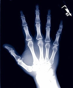
X-rays are high energy electromagnetic radiation. In other words, they are made of the same “stuff” as radiowaves, microwaves, infrared light, visible light, and UV rays, but are higher in energy. Because of their high energy, some X-rays can go right through the less dense parts of our bodies, such as skin and muscle, untouched, like visible light through a pane of glass.
These X-rays then reach a film, or detector, placed behind the patient and darken the image. However, the more dense parts of our body, our bones for example, absorb X-rays, preventing them from reaching the film or detector.
As we can see in this X-ray image of a hand, all of the x-rays which traveled through the air around the hand reached the film and darkened it. However, very few of the x-rays which had to travel through the thick layer of bone in the wrist reached the film, leaving a very light region in the image.
There is a clear contrast between bone and muscle tissue. The muscle tissue is less dense than bone, and so appears darker on the image.
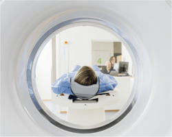
What is a CT scanner?![]()
In simple terms, it is a very powerful X-Ray machine. To imagine how a CT could be used to “look inside” the famous Enigma decoding machine we have to understand how X-Rays work.
A CT scan uses X-Rays to produce produces a three-dimensional image. Through a series of 2D X-ray images, taken along a ring perpendicular to the subject’s body, a 3D image of the inside of the subject is reconstructed using a computer.
CT scans are very helpful to doctors, as they give a complete picture of what is going on inside the body in three dimensions. In this very exciting, non-medical application a CT was used to uncover the secrets of Enigma. Because CT scans require a series of X-ray images to produce a complete picture, they emit much more ionizing radiation than a typical X-ray does. Whether it is a medical application or a fun research project like the one in the video below, we want to remind you that ionizing radiation, if left unchecked, carries serious risks.
Please be safe and join us at our X-Ray Safety Officer Courses or call our Free Information Service number 1800 – 263- 5803 to learn more about X-Rays and how to minimize your exposure risks.