Due to their high energy, x-rays and gamma radiation interact with materials differently than other lower-energy electromagnetic waves. Their unique properties have led to many applications in medicine, research, and industry for the purposes of imaging, treatment, and analysis. X-rays and gamma radiation are ionizing, which means they are capable of removing electrons from atoms and molecules. The International Agency on Research on Cancer (IARC) Monographs on the Identification of Carcinogenic Hazards to Humans[i] lists ionizing radiation as Group 1: Carcinogenic to humans.
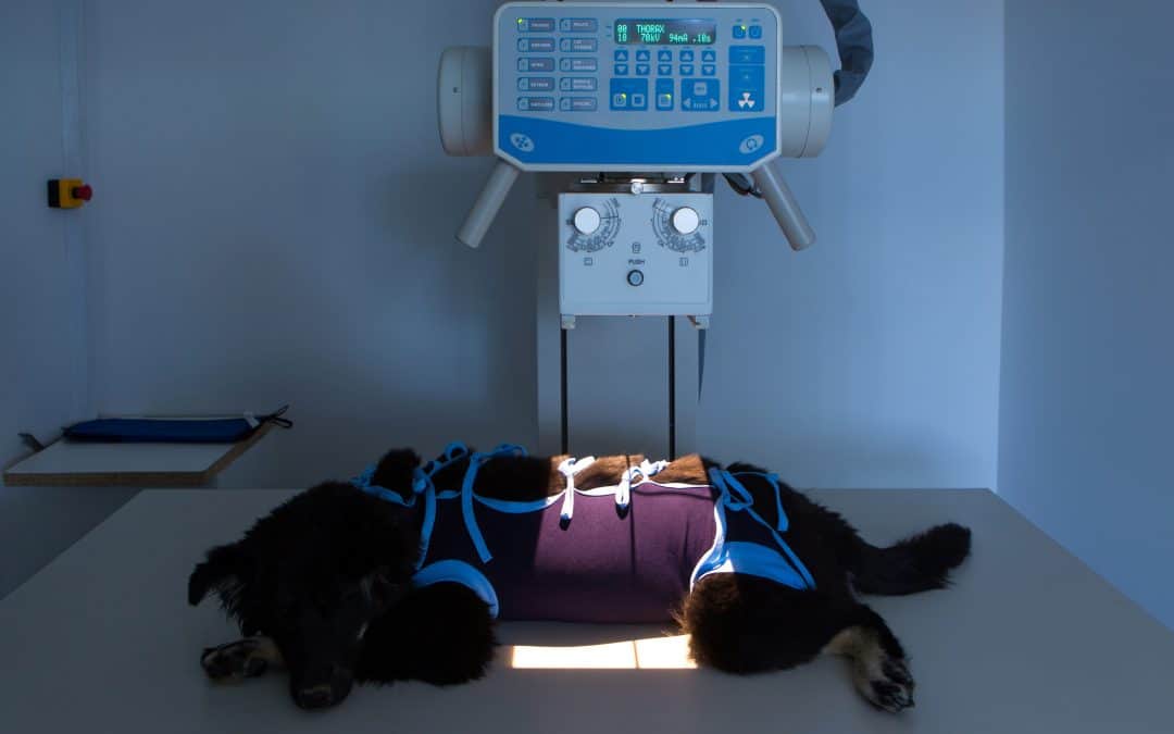
In veterinary practice, the animals being imaged cannot follow instructions to hold themselves still in a particular manner. As a result, x-ray imaging is done using restraint, sedation, and/or anesthesia. It is common for staff to manually restrain animals during x-ray examinations. In this blog post, we discuss radiation protection mindset and practices for veterinary radiography in face of the unique challenges due to the clientele.
Ionizing Radiation and Cancer Risk
Humans are regularly exposed to ionizing radiation from natural sources including solar and cosmic radiation; uranium, thorium, and their decay products found in the soil; ingesting radioisotopes found naturally in foods; and inhaling radon and radon decay products. We have evolved mechanisms to repair the damage to DNA from ionizing radiation and other carcinogens. Being exposed to a carcinogen does not automatically mean that a person will get cancer, but rather increase the person’s risk of getting cancer.
The rate by which being exposed to small doses of ionizing radiation over a long period increases the risk of getting cancer is not known. Countries which follow the recommendations of the International Commission on Radiological Protection (ICRP), including Canada, regulate the use of ionizing radiation under the precautionary assumption called the Linear No Threshold (LNT) Theory. This theory assumes that the risk for populations which receive low doses of radiation over time can be drawn from studies of populations of people who have received large doses in a short amount of time, such as the Japanese nuclear bomb survivors, and other research. It is therefore assumed that even a small dose of radiation can increase one’s risk of getting cancer.
Principles of Radiological Protection
No occupation is risk free. Workplaces in Canada have responsibilities under Occupational Health and Safety Acts to identify hazards and minimize risk to workers. In terms of radiological risks, the fundamental principles of radiological protection[ii] are:
- Justification: doing more good than harm;
- Optimization: keeping doses as low as reasonably achievable (ALARA); and
- Limitation: ensuring no person receives an unacceptably high dose.
These fundamental principles should be considered by employers when developing radiation protection expectations in the workplace as part of a management system committed to occupational health and safety. Documents such as Health Canada’s Radiation Protection in Veterinary Medicine (Safety Code 28)[iii], which is regulatory requirement in some jurisdictions in Canada, the ICRP’s Radiological Protection in Veterinary Practice (Publication 153)[iv], and the IAEA’s Radiation Protection and Safety in Veterinary Medicine (SRS No. 104)[v] can help support the development of a radiation protection program.
Hierarchy of Controls
Another OHS system which can be considered is the Hierarchy of Controls: an approach used across Canada and the United States to minimize or eliminate workplace hazards[vi],[vii],[viii]. This model helps to determine what practices are most effective in lowering risks. In order from most effective to least effective, those are:
- Elimination – removing the hazard altogether
- Substitution – using something else instead
- Engineering controls – isolating people from hazard
- Administrative controls – changing how people work
- PPE – Have people wear personal protective equipment
To lower the risk of cancer for veterinary workers, both the Principles of Radiological Protection and the Hierarchy of Controls can be applied for a more robust program. For example, when developing policies and procedures, thinking about the principle of justification can lead to questions such as, “In what cases is an x-ray mandatory? Are there other non-ionizing modalities such as ultrasound which could be of similar diagnostic value?”, which dovetail neatly into the controls of elimination and substitution.
Administrative Controls and PPE in Veterinary Practice
In the Hierarchy of Controls, Administrative Controls and PPE are shown to be the least effective methods of reducing workplace hazards. In veterinary practice where x-ray is deemed necessary, they cannot be ignored due to the possible requirement to have someone in the room with the veterinary patient.
For external sources of radiation, such as x-ray, the radiation protection principles of time, distance, and shielding are expected to be followed to help keep doses ALARA. It is expected that workers minimize the time spent at a task which exposes them to ionizing radiation, they maximize the distance from sources of radiation which decrease in strength when a person is farther away, and use appropriate shielding.
In terms of time, those manually restraining animals should not have any portion of their body in the beam used for imaging at any time. However, this in itself does not reduce worker dose to zero. Some portion of the x-ray beam used for imaging will scatter as the x-rays interact with the animal. Although lower in photon energy, this scatter is also ionizing and therefore carcinogenic. Studies have shown that there is measurable scatter radiation dose to restrainers[ix],[x],[xi]. Taking good quality images reduces dose to those giving restraint to the animal as it lowers the time of exposure by eliminating retakes.
While the use of PPE shielding also reduces dose to restrainers from scattered radiation[xii], studies have shown that many workers do not wear shielding during restraint or do so inconsistently or incorrectly[xiii],[xiv]. Inconsistent or incorrect wearing of PPE, combined with how manual restraint makes it difficult for workers to get adequate distance from the source of ionizing radiation (the veterinary patient causing the scatter) has led some veterinary workplaces to move to hands-free imaging techniques.
Hands-Free X-Ray Imaging
Hands-free imaging techniques in veterinary practice involve the use of administrative controls – changing how people work – to permit those who would typically be restraining the animal manually the ability to give themselves distance and/or remain behind shielding while in the room with the veterinary patient while obtaining images of diagnostic value.
Julia Bitan, a Canadian RVT, founded the Hands-Free X-rays Initiative[xv] to address concerns for radiation protection in veterinary medicine. For more information about how to implement hands-free procedures into your practice, please see our January 23, 2023 webinar[xvi] featuring an interview with Julia.
Concluding Message
It is important when creating a radiation protection program in veterinary practice to consider justification (making sure it is necessary), optimization (keeping doses as low as reasonably achievable), and limitation (keeping below dose limits). Controls such as elimination, substitution, and engineering controls should be implemented as part of any occupational health and safety program, including one for x-rays. In veterinary practice, where it is sometimes deemed necessary to have staff in the room with the patient, hands-free imaging techniques should be considered as a form of administrative control, along with all other administrative aspects such as proper record keeping and training. If PPE is being relied upon, remember workers are not to have any part of their body in the beam used for imaging, and they must correctly and consistently wear it.
[i] World Health Organization. (2023, July 19). IARC Monographs on the Identification of Carcinogenic Hazards to Humans. World Health Organization. https://monographs.iarc.who.int/list-of-classifications
[ii] ICRP.org. (n.d.). Fundamental principles of radiological protection. ICRPaedia. http://icrpaedia.org/Fundamental_Principles_of_Radiological_Protection
[iii] Radiation Protection in Veterinary Medicine – Recommended Safety Procedures for Installation and Use of Veterinary X-ray Equipment – Safety Code 28. (1991). Environmental Health Directorate Health Protection Branch. https://www.canada.ca/en/health-canada/services/environmental-workplace-health/reports-publications/radiation/radiation-protection-veterinary-medicine-recommended-safety-procedures-installation-use-veterinary-equipment-safety-code-28.html
[iv] Martinez, N., Bladel, L. V., Sovik, A., Balogh, L., Benoit, J., Davila, A., Dorling, S., Gambino, J., Natsuhori, M., Pentreath, R. J., Peremans, K., Randall, E., Roy, C., & Tanaka, I. (2022). Radiological protection in veterinary practice – ICRP Publication 153. SAGE. https://icrp.org/publication.asp?id=ICRP%20Publication%20153
[v] Radiation Protection and Safety in Veterinary Medicine – IAEA Safety Report Series 104. (2021). International Atomic Energy Agency. https://www.iaea.org/publications/13481/radiation-protection-and-safety-in-veterinary-medicine
[vi] Government of Canada, C. C. for O. H. and S. (2023, June 13). Hazard and risk – hierarchy of controls. Canadian Centre for Occupational Health and Safety. https://www.ccohs.ca/oshanswers/hsprograms/hazard/hierarchy_controls.html
[vii] Centers for Disease Control and Prevention. (2023, January 17). Hierarchy of controls. Centers for Disease Control and Prevention. https://www.cdc.gov/niosh/topics/hierarchy/default.html
[viii] Wikimedia Foundation. (2023, July 11). Hierarchy of hazard controls. Wikipedia. https://en.wikipedia.org/wiki/Hierarchy_of_hazard_controls
[ix] Barber, J., & McNulty, J. P. (2012). Investigation into scatter radiation dose levels received by a restrainer in small animal radiography. Journal of Small Animal Practice, 53(10), 578–585. https://doi.org/10.1111/j.1748-5827.2012.01257.x
[x] Belotta, A. F., Mayer, M. N., Waldner, C. L., Sidhu, N. P., Robinson, K. A., Carmalt, J. L., Freitas, F. P., & Koehncke, N. K. (2022). X-ray tube operators can be exposed to equal or higher scattered radiation doses to the hand as cassette holders during diagnostic radiographic procedures of the equine vertebral column and limbs. American Journal of Veterinary Research, 83(5), 412–418. https://doi.org/10.2460/ajvr.21.08.0134
[xi] Operator hand exposure to radiation during imaging procedures. (2012). Advances in Small Animal Medicine and Surgery, 25(3), 5–6. https://doi.org/10.1016/j.asams.2012.02.014
[xii] Mayer, M. N., Koehncke, N. K., Sidhu, N., Gallagher, T., & Waldner, C. L. (2019). Effect of full versus open-palm hand shielding on worker radiation dose during manual restraint for small animal radiography. Canadian Journal of Veterinary Research, 83(2), 154–158. https://www.ncbi.nlm.nih.gov/pmc/articles/PMC6450160/
[xiii] Mayer, M. N., Koehncke, N. K., Belotta, A. F., Cheveldae, I. T., & Waldner, C. L. (2017). Use of personal protective equipment in a radiology room at a Veterinary Teaching Hospital. Veterinary Radiology & Ultrasound, 59(2), 137–146. https://doi.org/10.1111/vru.12583
[xiv] Mayer, M. N., Koehncke, N. K., Taherian, A. C., & Waldner, C. L. (2019). Self-reported use of X-ray personal protective equipment by Saskatchewan Veterinary Workers. Journal of the American Veterinary Medical Association, 254(3), 409–417. https://doi.org/10.2460/javma.254.3.409
[xv] Hands-Free Veterinary Radiography Initiative. (n.d.). Hands-Free XRAYS. Hands-Free XRays. https://handsfreexrays.com/
[xvi] Radiation Safety Institute of Canada. (2023, June 28). Jan 23, 2023: Hands-Free Imaging in Veterinary Practice. Webinars – Radiation Safety Institute of Canada. https://radiationsafety.ca/services/webinars/#RSW12